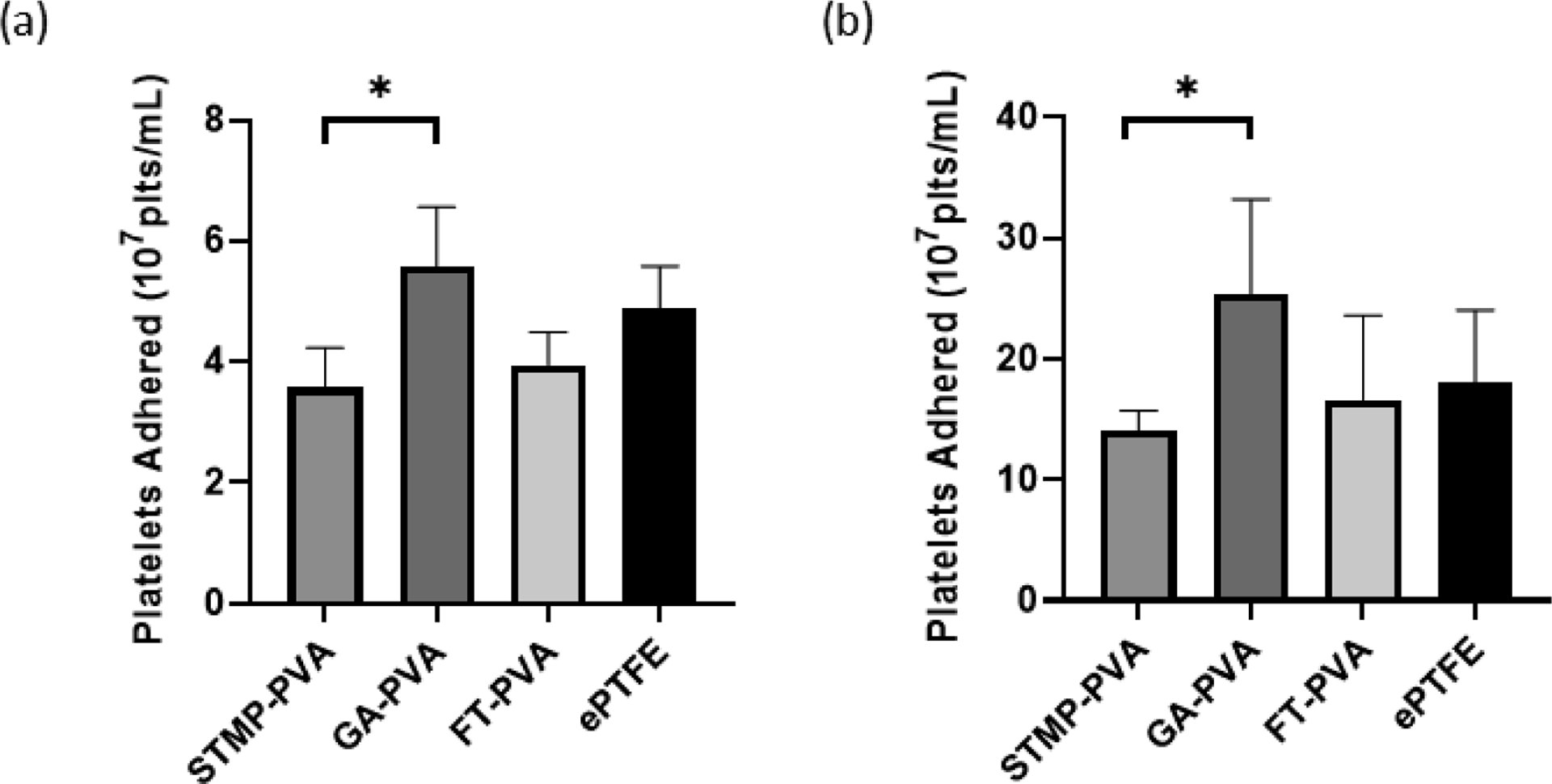
Figure 3.

Static platelet adhesion presented as the number of platelets present per mL of lysis buffer. (a) Isolated platelet adhesion onto crosslinked PVA biomaterials (n=3). (b) Static platelet adhesion onto crosslinked biomaterials in platelet-rich plasma (n=6). Data are shown as means ± SD. Statistical analyses were performed using a one-way ANOVA with Tukey’s post hoc testing. * indicate significant difference (p<0.05)
