
Figure 8. HMMR mediates autophagy repression by VPS35 D620N.
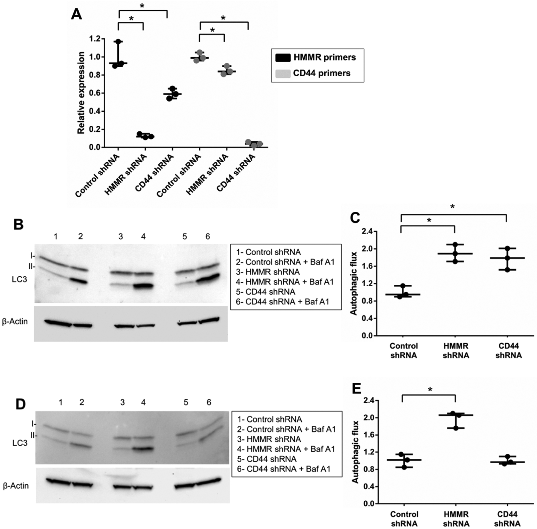
(A) Control, CD44 or HMMR expression was inhibited in differentiated SH-SY5Y cells neurons by lentivirus-delivered shRNA vectors. Transduced cells were selected with puromycin. Knockdown was validated by qPCR using the indicated primer sets and normalized to β-Actin transcript expression levels for each reaction. (B) Autophagic flux was examined in wild-type VPS35-expressing cells by western blot and quantified (C) following shRNA-mediated knockdown, treatment with or without bafilomycin A1 (Baf A1) and normalization of measurements to VPS35 WT shRNA control. (D) Autophagic flux was also assessed by western blot and quantified (E) in VPS35 D620N-expressing SH-SY5Y cells following shRNA transduction, Baf A1 treatment and normalization of measurements to VPS35 D620N shRNA control. Results from three independent experiments are shown (*p<0.05 versus indicated group, student’s t-test; error bars = SEM). Representative image groupings were obtained from single western blot membranes probed with multiple antibodies as indicated. β-Actin antibody was used as a loading control.