Fig. 1.
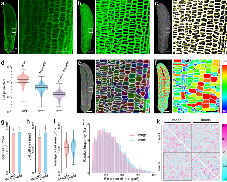
Recognition and qualification of Populus trichocarpa embryo cells by ImageJ and Imaris and their comparison. a Raw image of a Populus trichocarpa embryo captured by light sheet fluorescence microscopy (LSFM). b Compensation for the non-homogeneous fluorescent signal distribution in a using the ‘Plane Brightness Adjustment’ plugin. c Image of cell recognition and qualification by ImageJ software. d Quantification of cell area, perimeter, and Feret’s diameter from c. Boxplots represent mean, 25th, and 75th quartiles, whiskers represent minimum and maximum. n = 5845. e Image of cell recognition and qualification by Imaris software. f Heatmap of cell area calculated from e. The color scale represents the cell areas. g–j Comparison of values calculated by ImageJ and Imaris software. Statistical diagram of total cell number (g), total cell area (h), average cell area (i), and relative frequency of cell area. Boxplots represent mean, 25th, and 75th quartiles, whiskers represent minimum and maximum. n = 5845 and 6070. k Heatmap of correlation matrix analysis (Pearson, confidence interval = 95%) between cell areas (each cell area dataset is divided into 25 groups) calculated by ImageJ and Imaris software. In a–c, e, and f, panels on the right show enlarged images of areas on the left highlighted with white boxes; bar = 200 and 50 μm respectively

