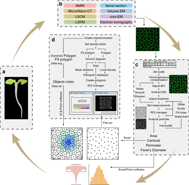
Fig. 2.
Flowchart of the procedure. a A plant sample (Arabidopsis seedling) prepared for analysis. b Basic raw imaging data for cellular outlines acquired by various 2D (two dimensional) and 3D imaging techniques used for this procedure; large-scale 3D images can be split into arbitrary 2D sections if needed. c Pre-processing, clarity adjustment, and parameter identification by ImageJ software. d Polygon creation, establishment of a Voronoï diagram, and object/cluster identification together with quantitative data generated and exported by SR-Tesseler software

