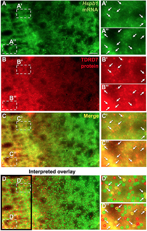
Figure 10.

Single-molecule fluorescence in situ hybridization (smFISH) coupled with immunostaining demonstrates TDRD7 protein to co-localize with Hspb1 mRNA in lens fiber cells. (A) Wild-type mouse lens at stage P15 was stained with complementary RNA probes specific to Hspb1 mRNA (green), (B) along with immunostaining for TDRD7 protein (red). (C) Merged image of the co-staining (yellow) and (D) analysis of significant co-localization of Hspb1 mRNA and TDRD7 protein using custom-written MATLAB program as described in Methods (colored open circles). (A’-D”) shows zoom-in of regions indicated by broken-line boxes in A–D. Arrows indicate co-localizing elements scored by the MATLAB analysis.
