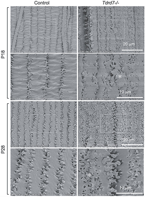
Figure 5.

Scanning electron microscopy demonstrates Tdrd7−/− lenses have abnormal fiber cell morphology. Scanning electron microscopy (SEM) was performed to visualize cortical fiber cells for control and Tdrd7−/− lenses at stages P18 and P28. While the control appeared normal, abnormal fiber cell morphology (asterisk) was observed in Tdrd7−/− lenses at both P18 and P28.
