
Figure 7.
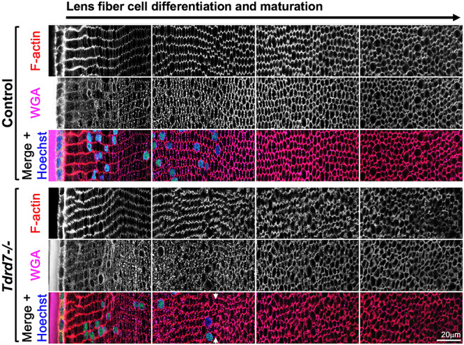
WGA and F-actin staining demonstrates abnormal membrane morphology and fiber cell organization specifically in fiber cells with nuclear degradation in Tdrd7−/− lenses. Control and Tdrd7−/− lens sections at mouse stage P15 were stained with wheat germ agglutinin (WGA) and phalloidin to visualize cellular membrane and F-actin, respectively. Hoechst stain was used to visualize DNA. Images from comparable areas representative of early to late fiber differentiation and maturation (left to right) are shown for both control and Tdrd7−/− lenses. While there is no discernable difference in young differentiation and maturing lens fiber cells at the lens periphery (left-most panel and left-half of the second panel) between control and Tdrd7−/− lenses, fiber cells showed abnormal WGA staining and abnormal fiber cell morphology in the Tdrd7−/− lens, coinciding with nuclear degradation (arrowheads), compared to control (second panel).