
Figure 2.
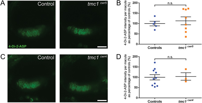
Tmc1 is not required for mechanotransduction in hair cells of the anterior macula as determined by 4-Di-2-ASP dye uptake. (A, C) Confocal micrographs of 4-Di-2-ASP fluorescence in hair cells of the anterior maculae of controls (left) and mutants (right). Controls are tmc1cwr5/+ (A) and wild-type larvae (C). (B, D) Plots of 4-Di-2-ASP fluorescence intensities of anterior maculae in tmc1 mutants. 6–8 dpf larvae were used. The control groups included both wild-type and heterozygous siblings. Each data point is the fluorescence intensity of one macula as a percentage of the average value of the control group. Mean ± SEM is displayed. (B) Intensity of tmc1cwr5 controls = 100% ± 11% (n = 5); intensity of tmc1cwr5 mutants = 113% ± 19% (n = 8). (D) Intensity of tmc1cwr4 controls = 100% ± 13% (n = 10); intensity of tmc1cwr4 mutants = 104% ± 18% (n = 3). Statistical significance determined by two-tailed unpaired t-test. Scale bar = 20 μm.