FIGURE 2.
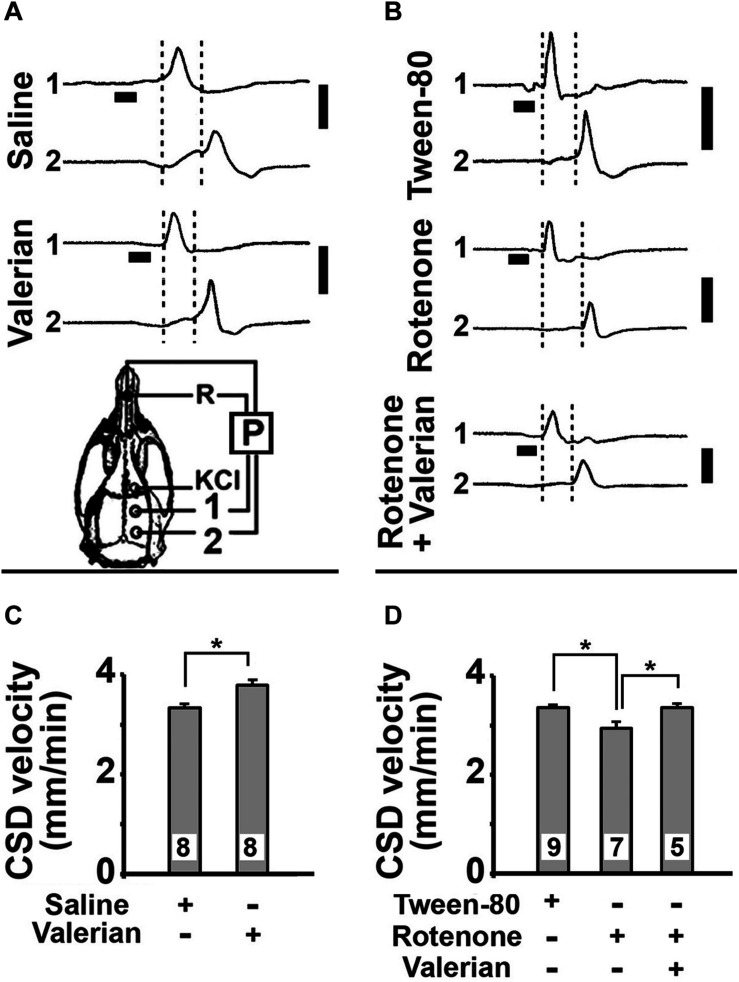
(A,B) Electrophysiological recordings (slow potential changes, P) of CSD in five rats that were treated per gavage with V. officinalis (250 mg/kg/d) or saline solution for 15 days, or with s.c. injections of Tween-80 (control group) or rotenone (10 mg/kg/d) for 7 days, or treated with s.c. injections of rotenone plus gavage with V. officinalis. The vertical solid bars at the right of the traces indicate 10 mV (negative upwards). The horizontal bars under the traces from the recording point 1 indicate the time (1 min) of application of the CSD-eliciting stimulus (a 2 mm diameter cotton ball soaked with 2% KCl solution) on the intact dura mater. Once elicited in the frontal cortex, CSD propagated and was recorded by the two cortical electrodes located at the parietal cortex (skull diagram, points 1 and 2). A third electrode of the same type was placed on the nasal bones and served as a common reference (R) for the recording electrodes. The vertical dashed lines delimited the latency for the CSD wave to cross the interelectrode distance. When compared with the corresponding controls, the latencies in the V. officinalis group and in the rotenone group were, respectively, shorter and longer. (C,D) CSD velocity of propagation in the 75–80-day-old rats of the five groups described in (A,B). Values are presented as the mean ± standard deviation of the 12 CSD measurements per rat, along the 4 h recording period, with the number of animals given at the bottom part of the bars. Asterisks indicate intergroup differences (p < 0.05; ANOVA plus the Holm-Sidak test).

