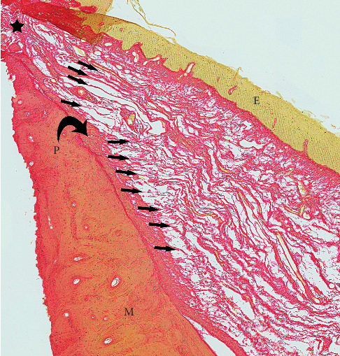
Figure 5.

Higher magnification of FOM fascia merging peripherally with periosteum. P: periosteum (curved black arrow). M: mandible. E: epithelium (FOM mucosal layer). Small arrows: FOM fascia merging with periosteum. Black star: lateral floor of mouth where FOM mucosa merges with gingival mucosa.
