Figure 9.

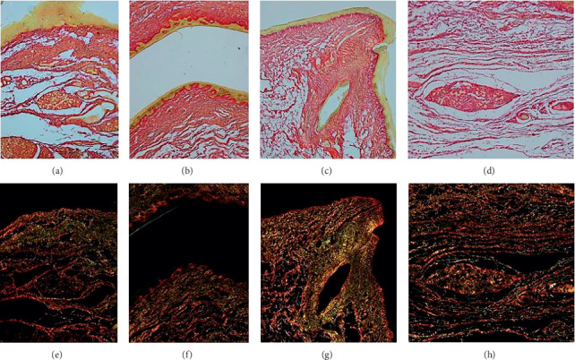
Collagen typing using polarized light microscopy (PSR). (a–d) Bright light microscopy: types I and III collagen stain red. (e–h) Corresponding images using polarized light microscopy: type III collagen highlights green.

Collagen typing using polarized light microscopy (PSR). (a–d) Bright light microscopy: types I and III collagen stain red. (e–h) Corresponding images using polarized light microscopy: type III collagen highlights green.