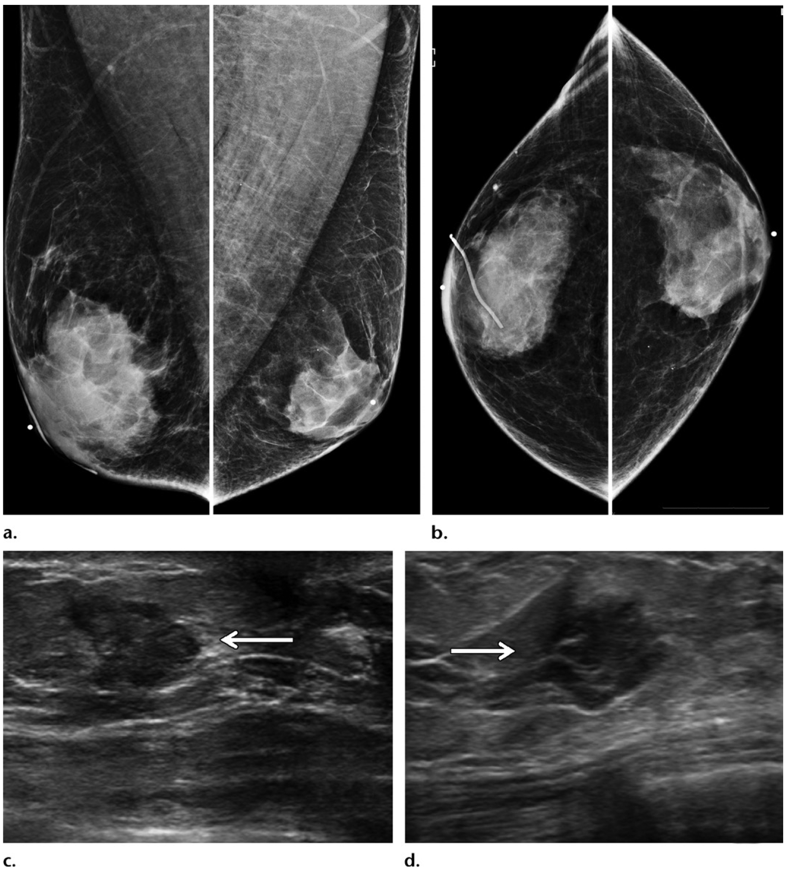
Figure 11.

Benefits of US in a case at high clinical suspicion for malignancy. A 50-year-old man of Ashkenazi Jewish ancestry who had bloody right nipple discharge and normal initial imaging workup underwent central duct excision at another institution, with surgical pathology results showing ductal carcinoma in situ with positive margins. This patient subsequently presented to our institution for further care. (a, b) Diagnostic MLO (a) and CC (b) mammographic views show the site of previous surgery in the right breast, as demarcated by a scar marker (a, b). Moderate bilateral gynecomastia was depicted as dense subareolar tissue in both breasts. No mass was seen at mammography. (c, d) Gray-scale US images show an irregular mass (arrow) in transverse (c) and sagittal (d) projections in the periareolar region of the left breast. A postoperative seroma was seen in the right breast (not shown). The patient underwent bilateral mastectomy, and surgical pathology results showed ER-positive, PR-positive, HER2-negative invasive ductal carcinoma in the left breast and ER-positive, PR-positive, HER2-negative ductal carcinoma in situ in the right breast.
