Figure 15.

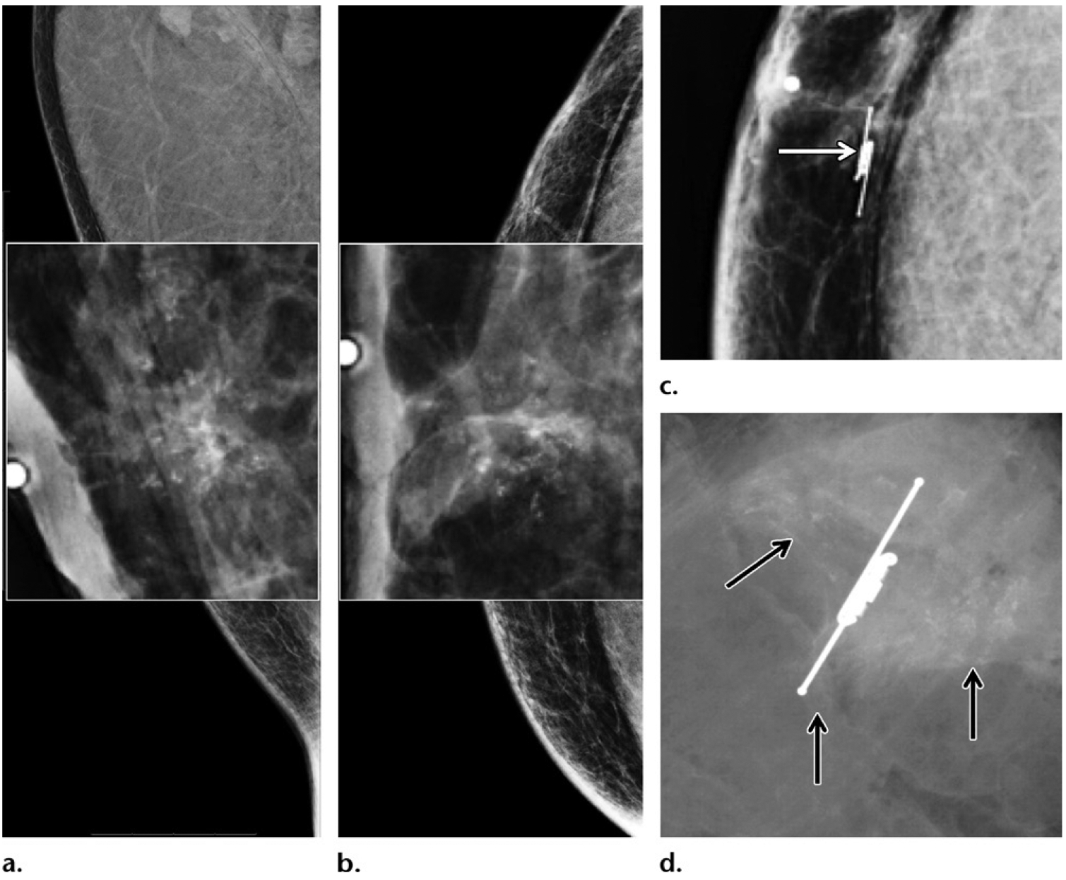
Screening and/or surveillance mammogram in a 54-year-old Ashkenazi Jewish man with a personal history of left breast cancer diagnosed 1 year prior and treated with mastectomy (same patient as in Fig 12), who tested positive for BARD1 mutation and had a family history of breast cancer (diagnosed in his father, two sisters, and paternal grandmother). (a, b) MLO (a) and CC (b) mammographic views of the contralateral right breast show new faint calcifications in the subareolar breast on the magnified views (at center). The patient underwent reflector placement for radar localization (Savi Scout; Cianna Medical, Aliso Viejo, Calif) of the calcifications prior to breast surgery. (c) Postprocedure mammogram in CC projection shows the calcifications demarcated by the reflector (arrow), to ensure removal. Right mastectomy was performed. (d) Radiograph of the surgical specimen confirmed presence of the reflector and the targeted calcifications (arrows). The histopathologic analysis showed ER-positive, PR-positive, HER2-negative ductal carcinoma in situ. This patient was therefore diagnosed with bilateral breast cancer early in the disease process on the basis of screening mammograms obtained within a 2-year span.
