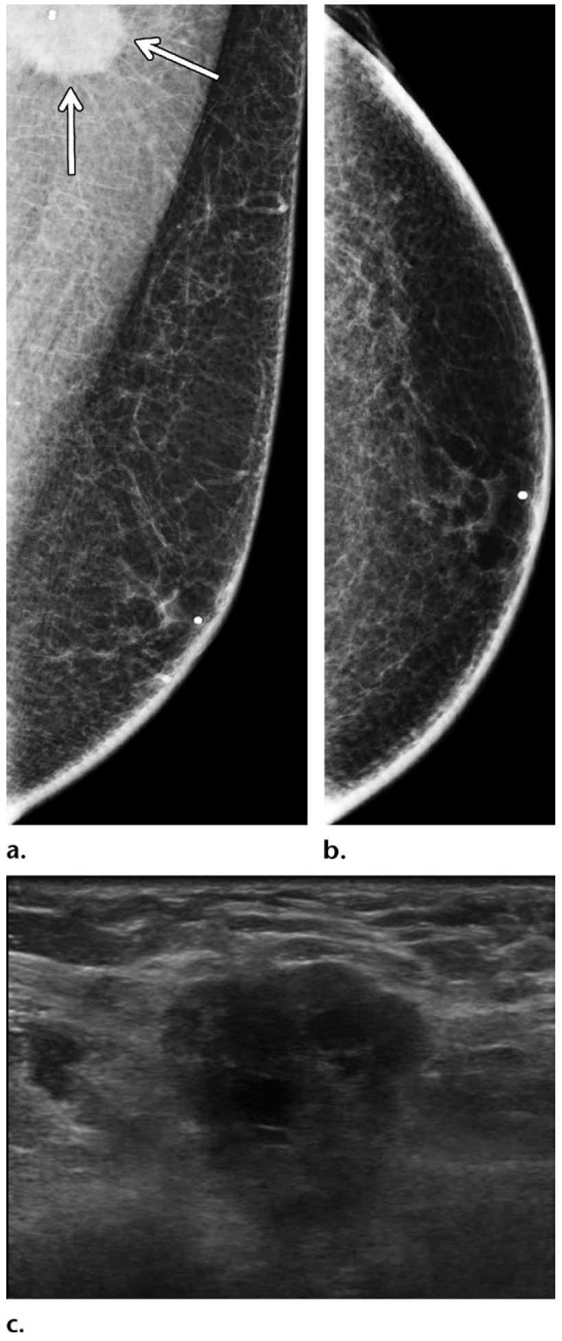
Figure 2.

Triple-negative breast cancer in a 54-year-old man who presented with an enlarging left axillary mass that was first palpated 3 years earlier. (a, b) Diagnostic MLO (a) and CC (b) mammographic views of the left breast show that an irregular mass (arrows on a) in the left axilla was only depicted on the MLO view. No other mass was depicted in either breast. (c) Gray-scale US image in sagittal projection shows a corresponding irregular mass with central areas of necrosis in the left axilla. The findings from histopathologic examination and testing of the specimen from core biopsy of the mass disclosed triple-negative (ER-negative, PR-negative, HER2-negative) invasive ductal carcinoma. The patient has a family history of breast cancer diagnosed in his mother when she was 52 years old; he has no known genetic mutation.
