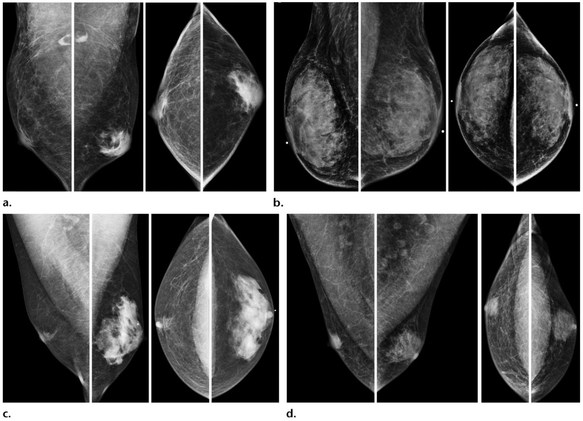
Figure 9.

Gynecomastia depicted as increased retroareolar fibroglandular tissue in four separate patients: Bilateral MLO and CC mammographic views of a 39-year-old man (a), a 75-year-old man (b), a 41-year-old man (c), and a 60-year-old man (d). Gynecomastia can often be asymmetric (a, c, d). Three subtypes of gynecomastia may be seen. Dendritic gynecomastia has a classic flame-shaped form, as seen in the right breast on c and the left breast on d. Nodular gynecomastia is depicted as a retroareolar mass in the right breast on d. Diffuse gynecomastia is seen as diffusely increased breast tissue bilaterally, mimicking female breasts, as depicted in both breasts on b. In this case (b), diffuse gynecomastia was a result of end-stage liver failure.
