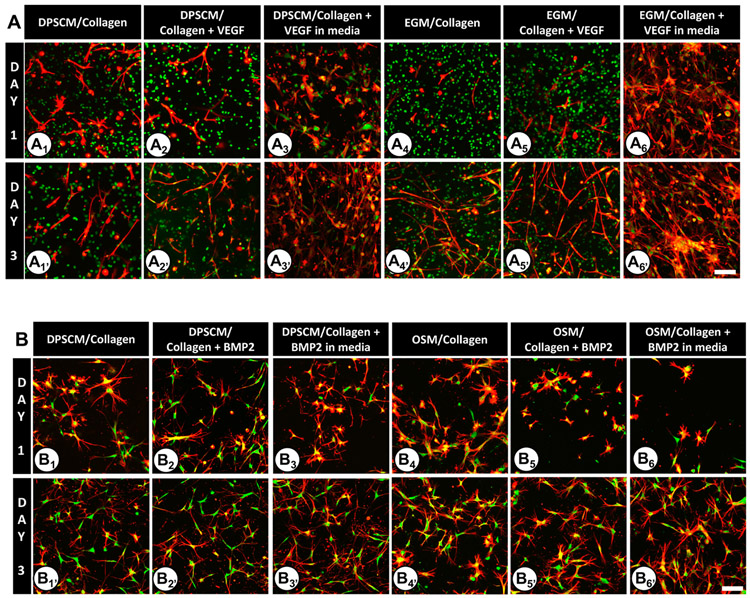
Figure 3.
DPSC adhesion and spreading within Oligomer 235 Pa collagen matrices under various conditions at Days 1 and 3 on the following matrices: (A1, A1′) DPSCM/collagen; (A2, A2′) DPSCM/collagen + VEGF; (A3, A3′) DPSCM/collagen + VEGF in media; (A4, A4′) EGM/collagen; (A5, A5′) EGM/collagen + VEGF; (A6, A6′) EGM/collagen + VEGF in media. DPSC adhesion and spreading within Oligomer 800 Pa collagen matrices under various conditions at Days 1 and 3 on the following matrices: (B1, B1′) DPSCM/collagen; (B2, B2′) DPSCM/collagen + BMP2; (B3, B3′) DPSCM/collagen + BMP2 in media; (B4, B4′) OSM/collagen; (B5, B5′) OSM/collagen + BMP2; (B6, B6′) OSM/collagen + BMP2 in media. For all images, the actin filaments are stained red with rhodamine–phalloidin (excitation and emission at 540/565 nm) and the nucleus is stained green using SYTO 13 (excitation and emission at 488/509 nm). Regions that appear yellow indicate the colocalization of actin and nucleus. Each experiment had three samples. Images are maximum intensity projection of Z-slices (~300 μm depth) captured using a confocal/2-photon Olympus FV1000 MPE system (Olympus America) with a XLUMPLFL20XW objective and 0.95 NA. Scale bar = 100 μm.

