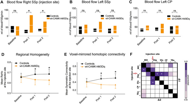
Figure 4.

Changes induced by activating hSyn-hM4Di in PVCre mice. (A) Neuronal firing rate at the right SSp DREADD injection site of the PVCre-hSyn-hM4Di mice before and after clozapine injection, indicating a steady increase in firing rate on DREADD activation. (B) Similar to figure A but for right caudoputamen, indicating no change in the neuronal firing rate after clozapine injection. (C) Comparison of blood flow (mL of blood/100 g/min) between controls (n = 7) and PVCre-hSyn-hM4Di (n = 13) mice over time (measured at the injection site). No significant differences observed between groups (repeated measures ANOVA: F1.4,17.9 = 2.45; P = 0.1). (D) Normalized average regional homogeneity (ReHo) change over time at the injection site. Repeated measures ANOVA indicates significant groups × scan time effect: F1,25 = 14.01; P = 0.001; asterisk indicates significant between-group independent samples post hoc t-test at ∆1 (t30 = −4.17; P = 10−5) and at ∆2 (t30 = −7.15; P = 10−6). (E) Normalized averaged symmetric connectivity over time at the injection site. Repeated measures ANOVA indicates significant groups × scan time effect: F1.2, 21.9 = 8.21; P = 0.007; asterisk indicates significant between-group independent samples post hoc t-test at ∆1 (t29 = −3.52; P = 0.001) and at ∆2 (t29 = −5.78; P = 10−5). (F) Seed-to-seed analysis indicates reduced FC (randomized permutation testing, P < 0.05, uncorrected) between controls (n = 13) and PVCre-hSyn-hM4Di (n = 19) mice. (G,H) Whole-brain connectome analysis shows a significant interhemispheric reduction between somatosensory cortices for ∆1 and ∆2 between PVCre-hSyn-hM4Di (n = 14) mice and controls (n = 13). Regions affected are as follows: SSp-m, primary somatosensory area, mouth; SSp-ul, primary somatosensory area, upper limb; SSs, supplementary somatosensory area; SSp-bdf, primary somatosensory area, barel field; SSp-n, primary somatosensory area, nose.
