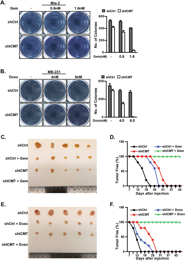
Fig. 2. Moderate ICMT knockdown enhances the effect of gemcitabine and doxorubicin in inhibiting the self-renewal and tumorigenic capacities of MiaPaCa2 and MDA-MB231 cells, respectively.
a MiaPaCa2 cells expressing either control or ICMT-targeting shRNA were cultured under the sphere forming conditions and treated with 0, 0.8, or 1.6 nM of gemcitabine as indicated. The sphere forming ability was assessed in three consecutive platings. The sphere images (left) were taken at the end of the third plating (3rd generation). The bar graph (right) presents the quantitation of sphere numbers from three technical repeats at the end of the 3rd plating; gray bar: control shRNA, white bar: ICMT-targeting shRNA. b Studies were performed as in a but with MDA-MB231 cells and that the chemotherapeutic agent used was doxorubicin at 0, 4, and 8 nM in combination with either control shRNA or that targeting ICMT. Data are presented as in a. For both panels, each sphere formation assay was performed with three technical repeats, and the experiments were repeated in three biological repeats with similar results. c, d MiaPaCa2 cells (80,000) expressing either control or ICMT-targeting shRNA were injected subcutaneously into contralateral flanks of NOD-SCID mice. Seven days after tumor implantation, the mice were divided into treatment and vehicle groups (n = 10 tumors each group); the treatment group received 150 mg/kg of gemcitabine twice a week. Tumor formation was monitored over the course of the study. At the end of the study, the mice were euthanized and the tumor excised and imaged (c). The percentage of tumor-free mice was plotted vs. treatment duration (d). e, f The same tumor formation study as shown in c and d was performed, except with MDA-MB231 cells and that the treatment group received doxorubicin at 1.5 mg/kg three times a week.

