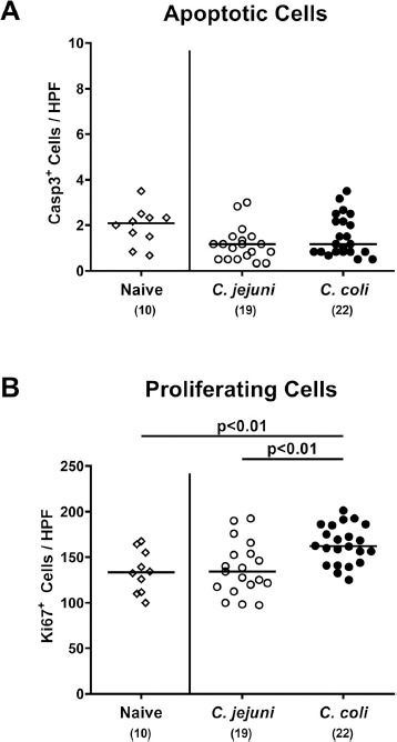
Fig. 6.

Colonic epithelial cell apoptosis and cell proliferation/regeneration in Campylobacter infected conventional mice. Conventionally colonized C57BL/6 mice were perorally infected with C. jejuni (open circles) or C. coli (closed circles) on day (d) 0 and d1 by gavage. On day 21 post-infection, the average numbers of colonic epithelial (A) apoptotic (Casp3+) and (B) proliferating (Ki67+) cells were assessed microscopically from six high power fields (HPF, 400 x magnification) per mouse in immunohistochemically stained colonic paraffin sections. Naive mice served as negative control animals (open diamonds). Medians (black bars), levels of significance (P-values) assessed by the Kruskal-Wallis test and Dunn’s post-correction and numbers of analyzed animals (in parentheses) are indicated. Data were pooled from four independent experiments
