Fig. 1.

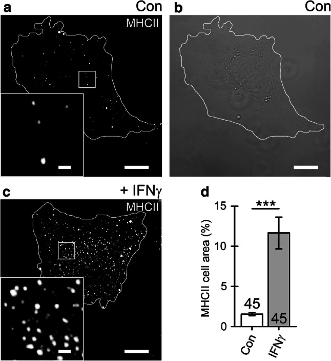
Cell treatment with IFNγ enhances the expression of MHCII that localize to vesicle-like structures in cultured rat astrocytes. a Confocal image of control (Con) astrocyte immunolabeled by anti-MHCII and secondary Alexa-546-conjugated antibody. b Differential interference contrast image of the same cell as in (a). c Confocal image of an astrocyte treated with IFNγ for 48 h. The white curve outlines the cell perimeter (a–c). Note numerous MHCII-positive vesicles in an IFNγ-treated astrocyte observed as bright fluorescent puncta. Insets display a magnified view of the MHCII-positive vesicles in control and IFNγ-treated cells. Scale bars: 10 μm (large images a–c) and 1 μm (insets a, c). d The relative proportion of MHCII-positive cell area (%; surface area of MHCII-positive pixels with fluorescence above 20% of maximal fluorescence) normalized to cell image area (surface area of all pixels delimited by the cell perimeter). The MHCII-positive cell area is substantially higher in IFNγ-treated astrocytes. The numbers at the bottom of the bars indicate the number of cell images analyzed. ***P < 0.001 versus control (Mann–Whitney U test)
