Fig. 9.

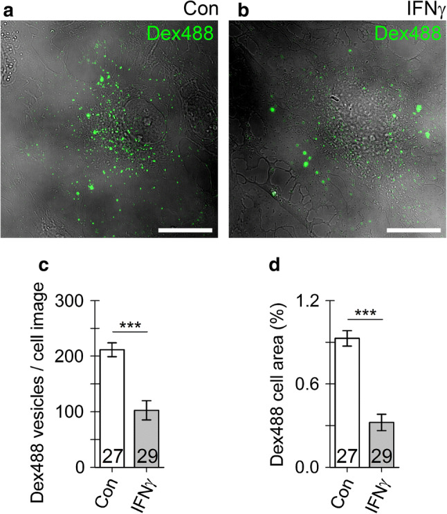
Fluid-phase endocytosis is suppressed in IFNγ-treated astrocytes. a, b Representative confocal images of an control astrocyte (a) and IFNγ-treated astrocytes (b) incubated for 3 h with 10 kDa dextran Alexa 488 conjugates (Dex488, green). Note individual Dex488-laden vesicles visible as bright fluorescent puncta (green). Scale bar: 20 µm. c, d Graphs displaying the number (mean ± SEM) of Dex488-laden vesicles per cell (c) and the relative proportion of Dex488-positive cell area normalized to cell image area (analogous to Fig. 1d) in controls (Con) and IFNγ-treated astrocytes. The numbers at the bottom of the bars indicate the number of cell images analyzed. ***P < 0.001 versus control (Mann–Whitney U test)
