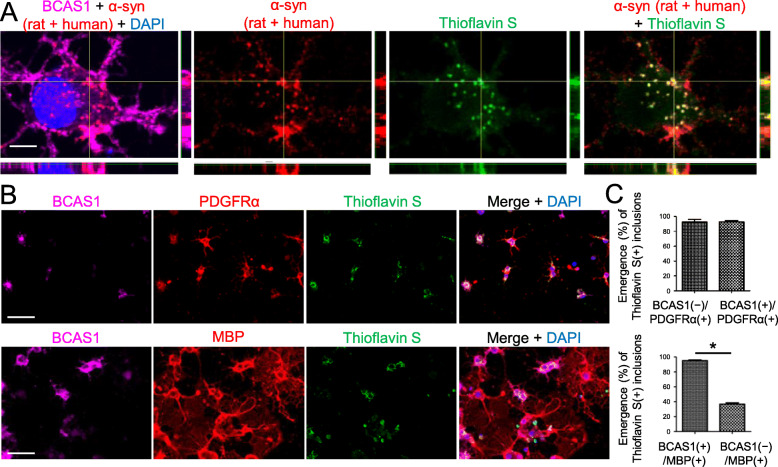
Fig. 3.
Extracellularly applied recombinant human α-syn PFFs induced cytoplasmic α-syn-immunoreactive inclusions in primary BCAS1(+) cell cultures. a Confocal images of BCAS1(+) cells, which were incubated with 1 μM α-syn PFFs for 24 h from day 4 after differentiation induction, showing the intracellular inclusions labeled with both anti-α-syn antibody and thioflavin S. Scale bar = 5 μm. b Immunostaining of oligodendroglial cells incubated with 1 μM α-syn PFFs for 24 h from days 3 (upper) and 4 (lower) after differentiation induction showing the ubiquitous development of thioflavin S-labeled inclusions in PDGFRα(+) cells and BCAS1(+) cells. In contrast, few BCAS1(−)/MBP(+) cells developed thioflavin S-labeled inclusions. Scale bar = 50 μm. c The percentages of oligodendroglial cells containing thioflavin S-labeled inclusions were compared between BCAS1(−)/PDGFRα(+) cells and BCAS1(+)/PDGFRα(+) cells (upper, performed on day 3), and between BCAS1(+)/MBP(+) cells and BCAS1(−)/MBP(+) cells (lower, performed on day 4). N = 4, respectively, independent culture, Mann–Whitney, p* < 0.05

