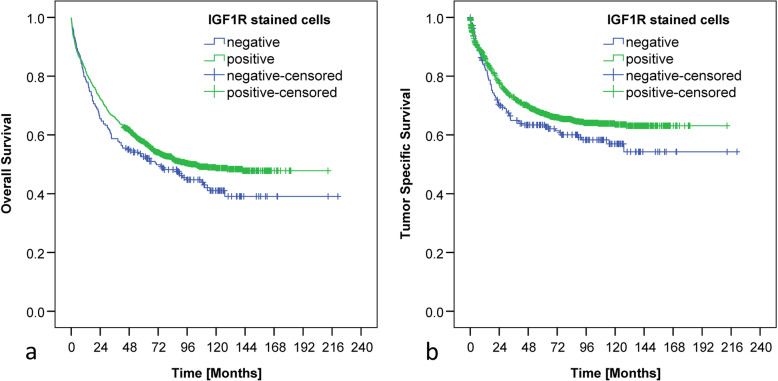
Fig. 3.
Kaplan-Meier curves based on the percentage of stained cells. Kaplan-Meier curves demonstrating correlations between IGF1 receptor expression in tumor cells and overall (a; p = 0.076) and tumor-specific (b; p = 0.076) survival based on the evaluation of the percentage of stained cells. CRCs with < 10% IGF1R positive tumor cells were declared as IGF1R negative and CRCs bearing ≥10% IGF1R positive tumor cells were classified as IGF1R positive. Neither the staining intensity nor the compartmental localization of the IGF1R were incorporated. Numbers at risk are provided below each Kaplan-Meier curve

