Figure 5.
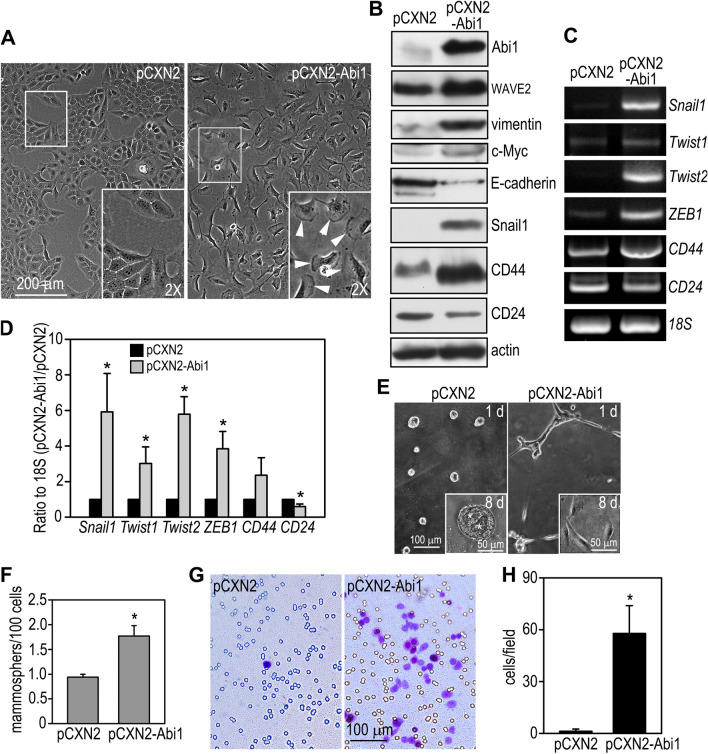
Overexpression of Abi1 in mammary epithelial cells induces the EMT and increases stem cell activity. (A) MCF10A human mammary epithelial cells were stably transfected with Abi1 (pCXN2-Abi1) or the vector alone (pCXN2). Live phase contrast micrographs show that overexpression of Abi1 converted MCF10A cells from an epithelial to a fibroblastic shape with prominent lamellipodia (arrowheads in the inset). (B) Control and Abi1-expressing MCF10A cells were subjected to immunoblot analysis. Actin served as a loading control. (C) RT-PCR showed that Abi1 overexpression increased the expression of EMT-inducing transcription factors and the ratio of CD44/CD24. (D) Ethidium bromide-stained gels were quantified by densitometry and plotted as a ratio to 18S. N = 3, *P < 0.05 vs cells transfected with pCXN2 only. (E) Live phase contrast micrographs show control and Abi1-overexpressing MCF10A cells cultured on Matrigel for 24 h and 8 days (insets). *Indicates cavity formation. (F) Abi1 overexpression increased mammosphere formation. N = 24, *P < 0.01. (G) Matrigel invasion assay showed increased cell migration to the bottom of the transwell after Abi1 overexpression. (H) Cells migrated to the bottom of the transwell were counted in 20 × field and plotted. N = 24, *P < 0.001.

