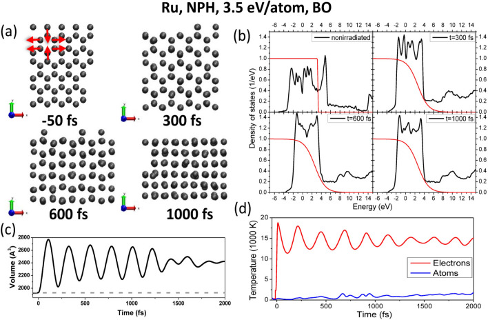
Figure 4.
(a) Atomic snapshots of NPH supercell of ruthenium irradiated with 3.5 eV/atom dose, modeled with XTANT-3 within BO approximation. Red arrows indicate the direction of atomic motion leading to nonthermal diffusionless phase transition. (b) Electronic DOS at the corresponding time instants; thin red lines depict electron distribution function. (c) Evolution of the volume of the supercell; the dashed line marks the volume of the supercell at ambient conditions. (d) Electronic and atomic temperatures evolution.

