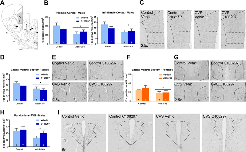
Figure 4: Fos immunoreactivity (Fos-ir) in the prelimbic and infralimbic divisions of the prefrontal cortex (PFC), lateral ventral septum (LSV) and parvicelullar division of the paraventricular nucleus of the hypothalamus (PVN).
Animals were subjected to adolescent CVS (adol) and concomitantly administered CORT108297 (30mg/Kg) or vehicle for 2 weeks starting at PND 46. Brains were collected 2h after the onset of FST, after 6 weeks of recovery from adol CVS. (A) Representative images from brain atlas showing the analyzed areas (Paxinos and Watson, 1998). (B-C) Fos-ir in the PFC of males and representative images. (D-E) Fos-ir in the LSV of males and representative images. (F-G) Fos-ir in the LSV of females and representative images. (H-I) Fos-ir in the PVN of males and representative images. *: significant result p < 0.05. Data are presented as mean ± s.e.m. n numbers are displayed in each bar.

