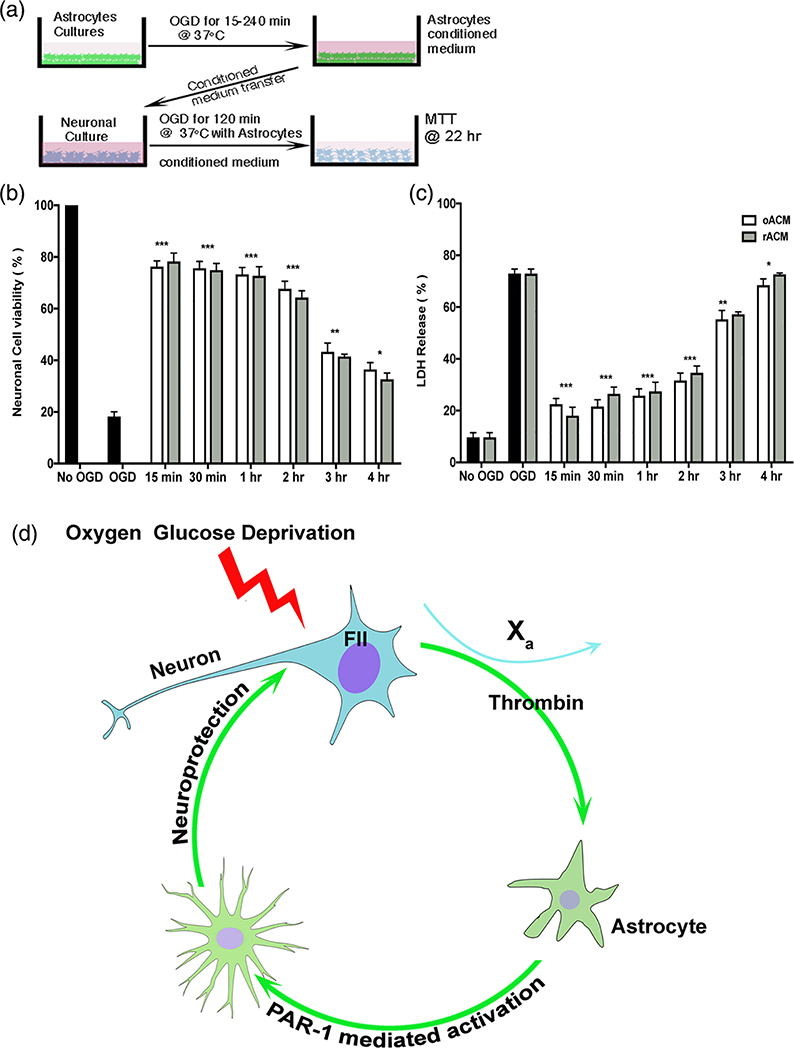
FIGURE 6.
Injured astrocyte media protects neurons undergoing OGD. (a) Experimental design to determine whether astrocyte conditioned media protects neurons during OGD. Astrocyte cultures were exposed to OGD for various times (15–240 min) followed by reperfusion for 1 hr. Astrocyte conditioned media after OGD (oACM) or reperfusion (rACM) was added to neuronal cultures prior to 2 hr OGD and 22 hr reperfusion. (b) Cell viability with methyl thiazolyl tetrazolium (MTT) and (c) cell death with the lactate dehydrogenase assay (LDH) were quantified as mean ± SEM. Neurons treated with oAMC showed significantly less cell death and increased cell viability compared to OGD alone. Neurons treated with rACM media were also protected. Two-way ANOVA p < .0001, Sidak’s correction for multiple comparisons, ***p < .0001. (d) Illustration of the hypothesis generated by the data presented here. Neurons exposed to injury (e.g., OGD) release prothrombin (coagulation factor II, or FII) that is converted to thrombin in presence of factor Xa. Thrombin activates nearby astrocytes via PAR1. Activated astrocytes release paracrine neuroprotective factors into the surrounding environment thereby protecting neurons from further injury. OGD, oxygen-glucose deprivation

