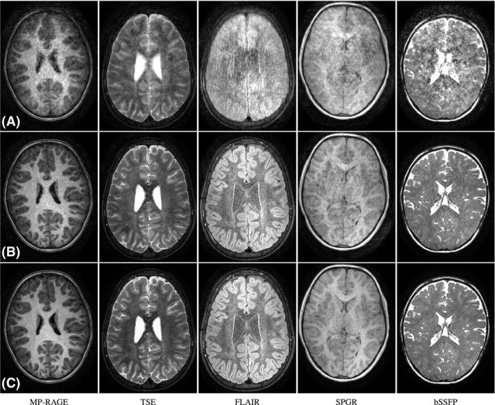
Figure 8.

Reconstruction results for pediatric cases with largest intra‐scan degradations. A, Uncorrected; B, motion‐corrected; and C, motion‐corrected and regularized outlier segment rejection reconstructions. From left to right, results for the main families of sequences for structural brain imaging
