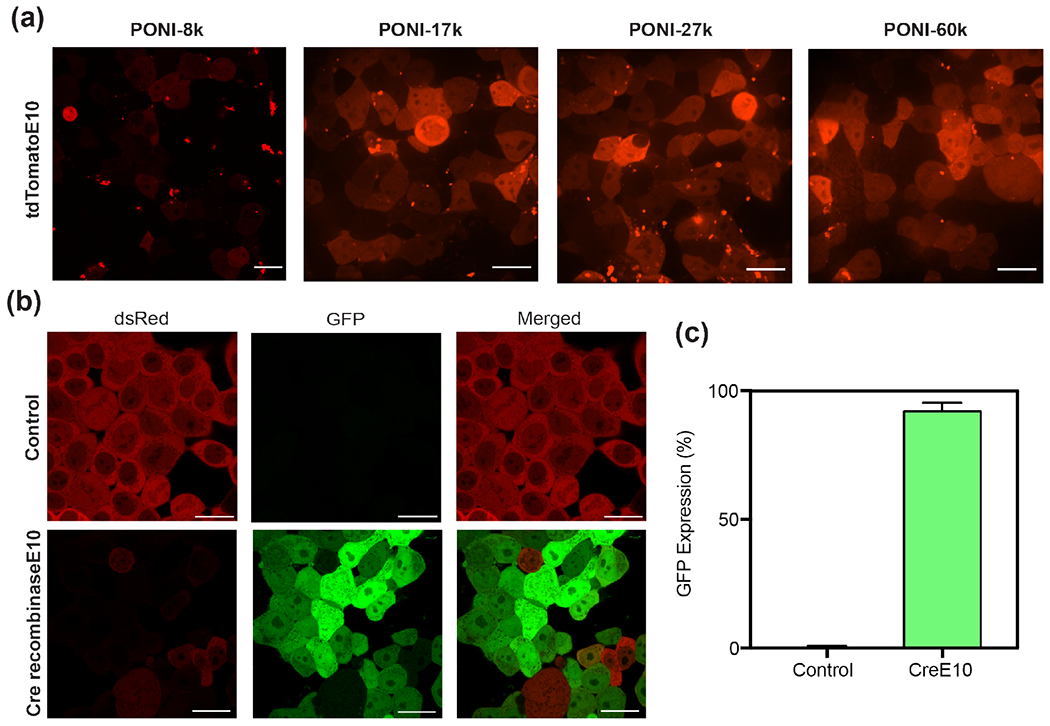
Figure 7.

Representative confocal images (40x) of (a) tdTomato-E10 delivery to HEK-293T using PONI-27k after 24h PPNC incubation, and (b) Cre-reporter HEK-293T cells after 24h PPNC incubation and an additional 24h growth period. Scale bars=20 μm. (c) Percentage of GFP+ cells, determined by analysis through ImageJ. Additional confocal images used for analysis are available in Figure S17.
