
Figure 3. EGFR L718Q and L718V mutations confer resistance to osimertinib in human lung cancer cells.
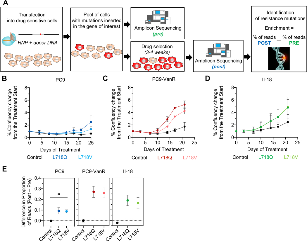
A. Schematic workflow of the CRISPR experiment (see Supplementary Methods). B-D. Cell growth of CRISPR-edited-PC9 (blue), PC9-VanR (red), and II-18 (green, lines superimposed) cells during osimertinib treatment as measured by Cell Metric. E. Histograms showing the difference in the proportion of reads for the indicated codon in the corresponding sample before (pre) and after osimertinib selection (post). As a reference, the mean of the top 5 indels found in the control sample is shown. The dashed line is the background threshold. Error bars show SEM for three biological replicates for PC9 and two for PC9-VanR and II-18 for B-D and E. Two-sided t test, * p<0.05.