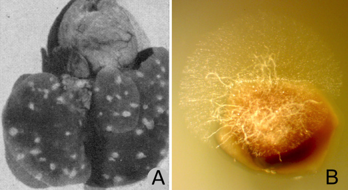
Fig 1. Small-mammal lung tissues showing fungal growth.
(A) Lung from Peromyscus sp. collected in the vicinity of San Carlos, Arizona, with fungal lesions believed to be Emmonsia parva (Blastomyces parvus), reproduced from Emmons and Ashburn [21]. (B) Fungal hyphae in lung tissue from apparently healthy Dipodomys heermanni collected in Kern County, California, (MVZ:239394) from which E. parva was recovered in pure culture. The lung fragment shown was incubated for 48 hours on water agar with tetracycline (10 mg/ml) and chloramphenicol (50 mg/ml). The fungal growth shown in B is typical for small-mammal lungs we have examined from diverse species. Segments of any given lung plated on growth medium will often result in growth of multiple fungal species. The image shown in A is from Public Health Reports (volume 57) and is in the public domain.

