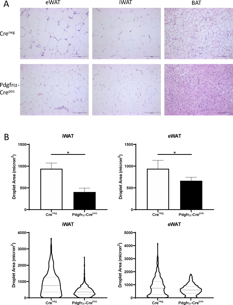
Fig 5. Adipose tissue pathology in mice on HFD without Ahr knockout (Creneg) or with Pdgfrα-Cre Ahr knockout (Pdgfrα-Crepos).
A. Shown are representative examples of Creneg and Pdgfrα-Crepos mice on HFD for eWAT, iWAT, and BAT. Scale bars are 100 micrometers. B. Quantitation of adipocyte sizes in eWAT and iWAT fat depots of Creneg or Pdgfrα-Crepos mice on HFD. Quantitation was performed using the Adiposoft program in ImageJ. The upper panels represent the mean and the error bars are standard deviation. Unpaired T-test, *p<0.05. The lower panels contain descriptive graphs to show the 25th, 50th, and 75th quartiles to illustrate the large differences ins droplet sizes between the groups.

