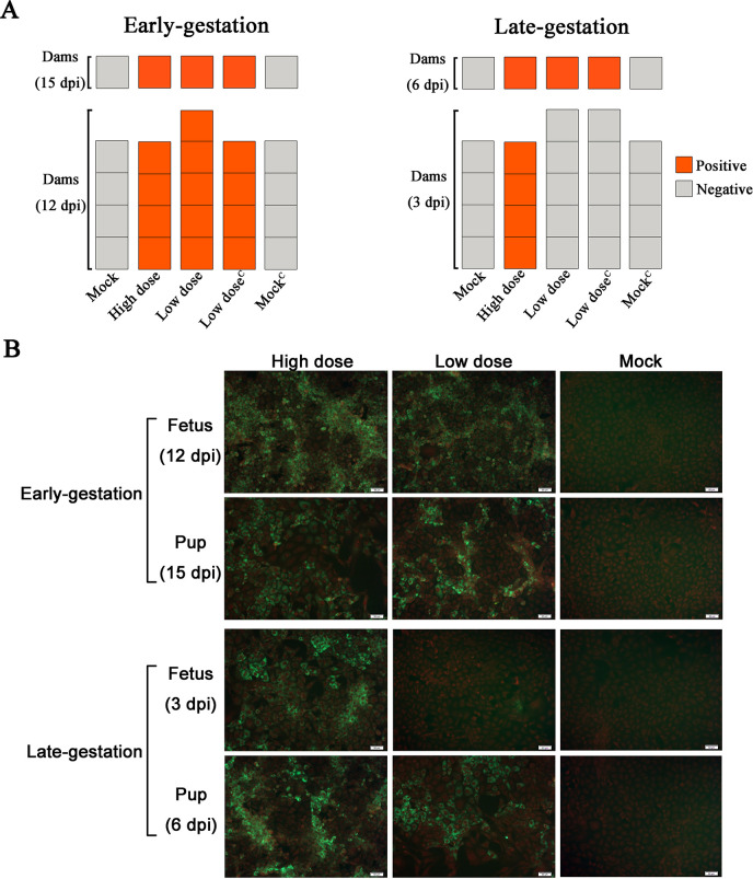
Fig 4. Anti-SFTSV total antibodies in dams and the infectious virus in the viscera of fetuses and pups.
(A) Anti-SFTSV total antibodies were detected in dams by ELISA. Each panel represents a dam, orange is positive for Anti-SFTSV total antibodies and grey is negative. The upper row of panels represents the dams after normal delivery. (B) Detection of infectious SFTSV in the viscera of fetuses and pups. Red indicates the nucleus, and the cytoplasm was labeled with 0.5% Evans blue. Green indicates the distribution of infectious SFTSV in monolayer Vero cells. Images (200×) of cells, scale bars, 50 μm.

