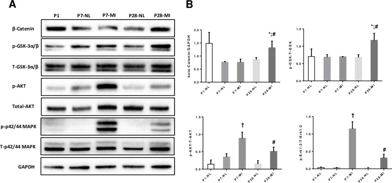
Fig 6. Western blotting. LAD ligation at P1 activates key cell proliferation signaling pathways and downstream components.
The expression of β-catenin, phosphorylated (Pho-) GSK 3α/β, total GSK 3α/β, Pho-Akt, total Akt, Pho-p42/44 MAPK, and total p42/44 MAPK was evaluated via Western blot. GAPDH levels were also evaluated to confirm equal loading. (A) Representative Western blotting of hearts from each time point/group; (B) Compiled Wetern blotting data, n = 3 hearts each bar. T-test (2 tails) with Bonferroni correction. *, p<0.05 between P28-MI vs P28-NL; #, p < 0.05 vs P7-MI; †, p<0.05 vs P7-NL. Total protein blots were the same original blots stripped and reprobed.

