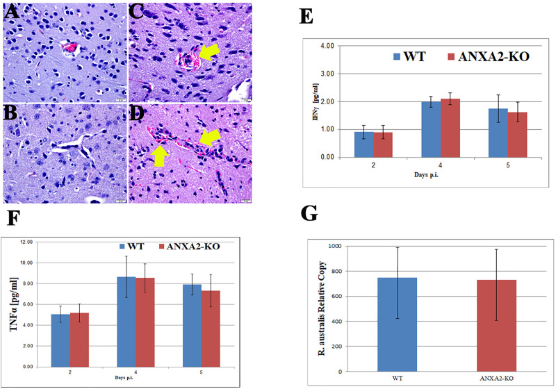
Fig 2.
Representative H&E staining of brain sections from WT (A&B) and ANXA2-KO (C&D) 5 days post-R. australis infection. Perivascular hemorrhage (yellow arrow) can be observed in infected ANXA2-KO group but not infected WT group. TNFα (E) and IFNγ (F) concentrations in serum at 2,4,5 days post-R. australis infection. Relative R. australis DNA copies (G) extracted from the brain of WT and ANXA2-KO mice quantified by rt-qPCR. No significant difference was found. Error bar stands for standard deviation. Scale bar: 20 μm.

