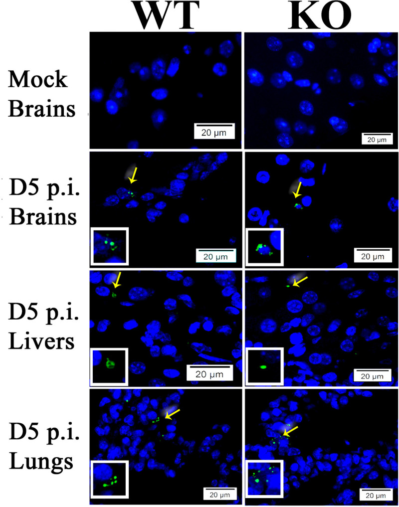
Fig 3.

Representative IF staining of SFG rickettsiae (green) in livers, brains, and lungs from WT and ANXA2-KO mice with nuclei of host cells counter-stained with DAPI (blue). The areas indicated by the arrows are enlarged and distinguish rickettsial (green) staining (boxed inserts). Scale bars, 20 μm.
