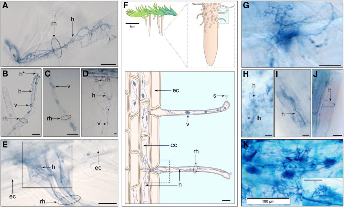
Fig. 1.
Fine root endophyte (FRE) hyphae with vesicles and fan-like structures in mature Lycopodiella inundata sporophytes. Root hair cells (a–e), showing examples of fine hyphae and a–c intercalary vesicles or hyphal swellings ranging 2–10 μm. Some hyphae were seen entering through the root hair tip (labelled h* in b). e Two adjacent root hair cells with bundles of FRE strands twisting and branching throughout, here colonizing cortical cells but skipping epidermal cells. f Schematic sketch of a Lycopodiella inundata plant illustrating a root, root hair cells, fine hyphae, vesicle, spores and epidermal and cortical cells (expanded shaded box on bottom). The dotted square in e highlights a root hair position between epidermal cells illustrated in the equivalent dotted box of the sketch. Fan-like FRE (g) were observed branching and twisting throughout the root. h An individual cortical and i, j epidermal cells with FRE. k Previously published FRE and arbuscules (inset) in Trifolium subterraneum root (adapted from Orchard et al. 2017aNew Phytologist with permission). All micrographs are acidified Sheaffer blue ink. Labels: ‘rh’ root hair; ‘h’ fine hyphae; ‘v’ intercalary vesicle; ‘s’ spore; ‘cc’ cortical cell; ‘ec’ epidermal cell. All scale bars 20 μm, unless detailed otherwise

