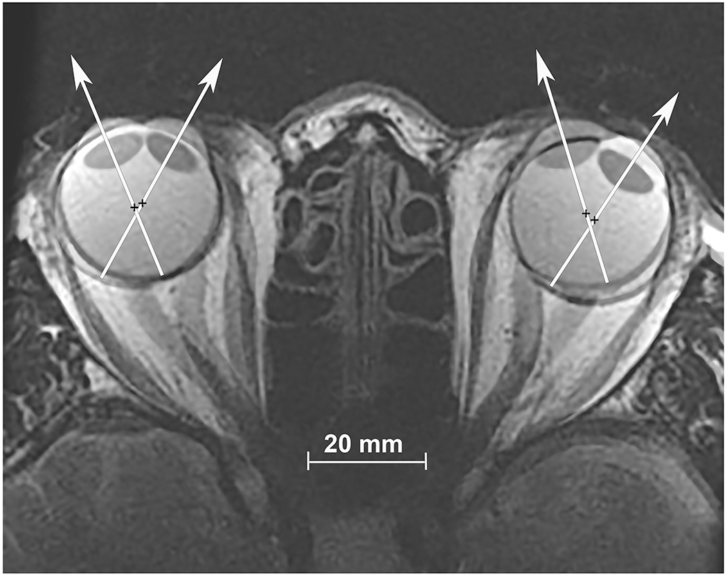
Fig. 1.

Axial, T2-weighted MRI obtained using surface coils in a normal subject fixating in right gaze, and superimposed at partial opacity on the image separately obtained in left gaze. Visual axes are depicted by superimposed white arrows, and rotational center by black crosses. Note the globe translation during this large horizontal rotation of each eye.
