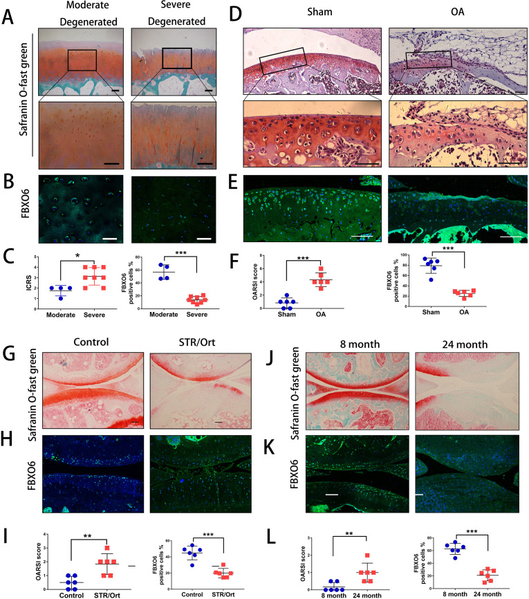
Figure 1.
Decreased expression of FBXO6 in chondrocytes during osteoarthritis (AO) development. (A) Safranin O-Fast Green staining in human moderate and severe degenerated cartilage. Insets indicate the regions shown in the enlarged images (down). (B) Immunostaining for FBXO6 in moderate and severe degenerated cartilage. (C) ICRS (International Cartilage Repair Society) scores and quantification of FBXO6-positive cells in moderate and severe degenerated cartilage. (D) Safranin O-Fast Green staining in mouse sham and OA tibial cartilage. Insets indicate the regions shown in the enlarged images (down). (E) Immunostaining for FBXO6 in knee joint of sham and OA mouse. (F) Osteoarthritis Research Society International (OARSI) histological scores and quantification of FBXO6-positive cells in knee joint of sham and OA mouse. (G) Safranin O-Fast Green staining in control and SRT/Ort mouse joints. (H) Immunostaining for FBXO6 in control and SRT/Ort mouse joints. (I) OARSI histological scores and quantification of FBXO6-positive cells in knee joint of control and SRT/Ort mouse. (J) Safranin O-Fast Green staining in 8-month and 24-month mouse joints. (K) Immunostaining for FBXO6 in 8-month and 24-month mouse joints. (L) OARSI histological scores and quantification of FBXO6-positive cells in 8-month and 24-month mouse joints. (Scale bars: 100 µm. Data are expressed as mean±SD. **p<0.01; ***p<0.001.)

