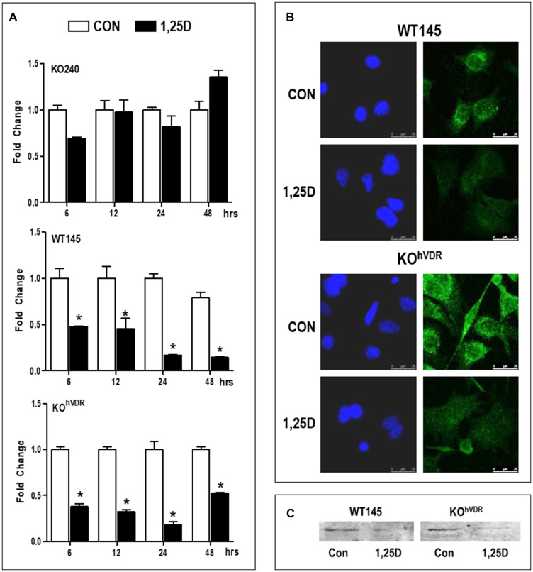
Figure 1. VDR is required for 1,25D3 mediated down-regulation of Has2 mRNA and protein.
(A) RNA was isolated from KO240, WT145 and KOhVDR cells treated with 100 nM 1,25D3 for 6, 12, 24, or 48 hours. Has2 mRNA in control and 1,25D3 treated samples was assessed by the ΔΔCt method and values were normalized against 18S and expressed as fold change (1,25D3 vs control) for each cell line. Bars represent mean ± standard deviation, * p < 0.05 control vs 1,25D3 treated at each time point as evaluated by Student’s t test. (B) Immunofluorescence for HAS2 (green) in WT145 and KOhVDR cells treated with 100 nM 1,25D3 or vehicle for 48 hours. Nuclei were stained with DAPI (blue). Images were acquired on a Leica DMI6000 microscope with a TCS SP5 confocal laser scanner using Leica Application Suite software. (C) Lysates from WT145 and KOhVDR cells treated with 100 nM 1,25D3 for 48 hours were blotted with antibodies against HAS2.

