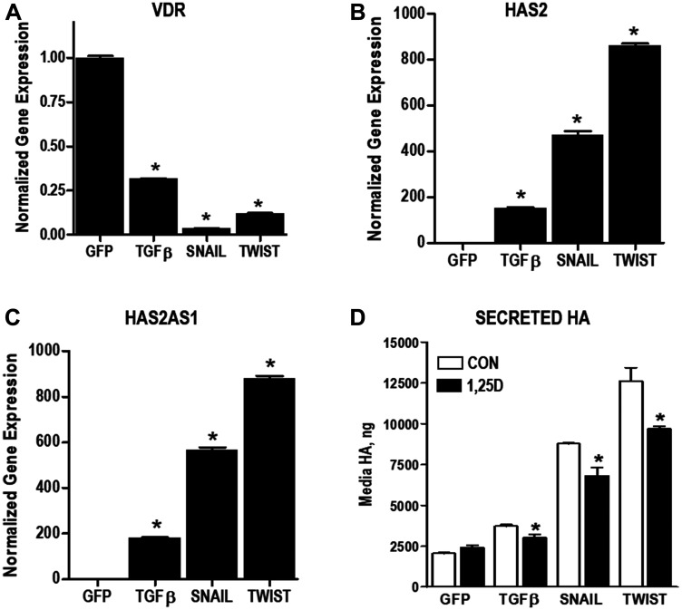
Figure 6. Changes in VDR and HA pathway during EMT.
(A–C) RNA was isolated from immortalized human mammary epithelial (HMLE) cells stably expressing GFP (control), TGFβ, SNAIL or TWIST. Expression of VDR (A), HAS2 (B) and HAS2AS1(C) were evaluated by RT-qPCR, normalized to 18S expression and expressed relative to GFP control cell line. Bars represent mean ± standard deviation of at 3 independent samples analyzed in triplicate. * p < 0.05 vs expression in HMLE cells as determined by one-way ANOVA and Dunnett’s post hoc test. (D) Secreted HA was evaluated in conditioned media of HMLE cells stably expressing GFP, TGFβ, SNAIL or TWIST by ELISA. * p < 0.05, 1,25D3 treated vs control for each cell line as evaluated by Student’s t test.

