Fig. 3.
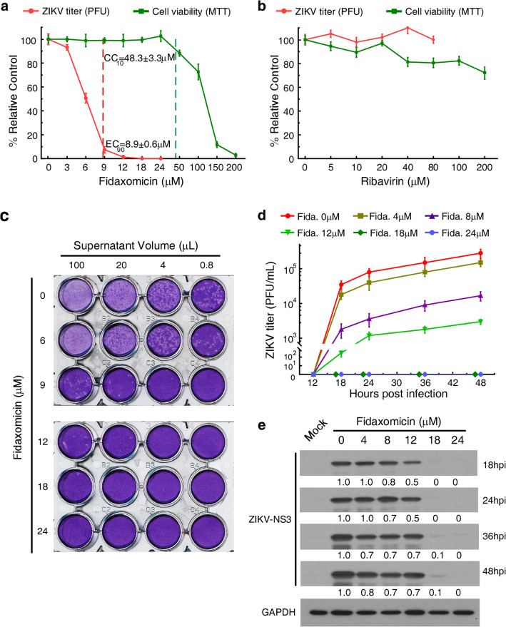
Fidaxomicin blocks ZIKV infection in vitro. a Antiviral and cytotoxicity spectrum of fidaxomicin in SNB19 cells. ZIKV (ZG-01) titer (the red line) was quantified by plaque assay, and cell viability (the green line) was detected by 3-(4,5-dimethylthiazol-2-yl)-2,5-diphenyltetrazolium bromide (MTT) assay. The values of EC90 and the CC10 are marked respectively. The data are the mean values ± standard deviation from triplicate experiments. b Antiviral and cytotoxicity spectrum of ribavirin in SNB19 cell line. ZIKV (ZG-01) titer (the red line) was quantified by plaque assay, and cell viability (the green line) was detected by MTT assay. The data are the mean values ± standard deviation from three independent experiments performed in triplicate. c Representative images of plaque assay. The ZIKV (ZG-01) titer in the supernatant was detected by plaque assay on new monolayers of Vero 76 cells. The supernatant was obtained from SNB19 cells culture treated with fidaxomicin at indicated concentrations at 48 h post-infection (hpi). d The anti-ZIKV activity of fidaxomicin in SNB19 cells. Vero cells were infected with the supernatant obtained from SNB19 cells culture treated by fidaxomicin at indicated concentrations, and the ZIKV (ZG-01) titers were detected by plaque assay at indicated hpi. Data are representative of three independent experiments performed. e Western blotting analysis of protein expression of ZIKV NS3 in the cell lysates of SNB19 for the anti-ZIKV activity of fidaxomicin at indicated hpi

