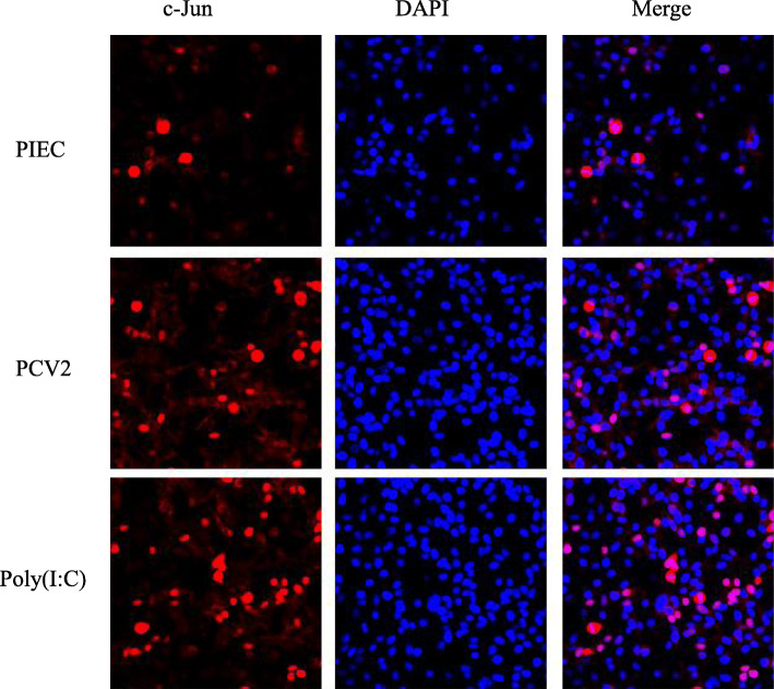
Fig. 6.
Nuclear translocation of JNK after PCV2 infection in PIECs. PIECs were infected with PCV2 (MOI = 0.5) for 1 h and then cultured for 24 h. Fluorescence confocal microscopy was used to measure the cellular localization of c-Jun in PCV2-infected PIECs. The localization of c-Jun (red) was observed with a fluorescence microscope using immunofluorescence staining with anti-c-Jun and Alexa Fluor®647 conjugate anti-rabbit IgG. Nuclei were stained with DAPI. The uninfected PIEC was used as a negative control and Poly(I:C) treatment was used as a positive control. Bar = 20 μm

