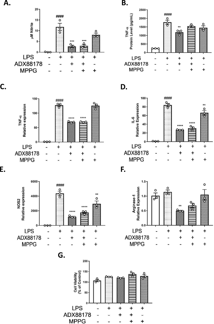
Fig. 4. Effect of mGluR4 antagonist, MPPG, in LPS-stimulated mouse primary microglia.
Primary microglia cells were pre-treated with group III antagonist, MPPG, for 1 hour prior to addition of ADX88178 for 30 minutes followed by stimulation with or without LPS (20ng/mL) for 24 hours. (A-B) LPS stimulation resulted in a significant increase in nitrite production and TNF-α release (####P<0.0001 vs. control for both). Pre-treatment with 20mM of ADX88178 significantly reduced LPS-induced nitrite production (***P<0.001 vs. LPS) and TNF-α levels (**P<0.001 vs. LPS). However, MPPG failed to inhibit anti-inflammatory actions of ADX88178 as measured by NO levels and TNF-α protein levels. (C-E) Expression of pro-inflammatory mediators in primary microglia was measured at 6h. LPS stimulation of primary microglia significantly increased expression of TNF-α, IL-6, and NOS2 (####P<0.0001 vs. control for all three genes). ADX88178 pre-treatment resulted in a significant downregulation of LPS-induced levels of TNF-α, IL-1β, and NOS-2 (****P<0.0001 vs. LPS for all three genes). However, MPPG failed to inhibit the anti-inflammatory actions of ADX88178 as measured by these pro-inflammatory gene expressions. (F) There were no significant changes in the gene expression of anti-inflammatory mediator Arginase-1. (G) There was no cytotoxicity at the concentrations of drugs used as measured by MTT assay. All values are expressed as mean ± S.E.M. for at least three independent experiments. Data were analyzed using one-way analysis of variance (ANOVA) for multiple comparisons with post hoc Tukey’s test. ####P<0.0001 in comparison with control; **P<0.01, ***P<0.001, ****P<0.0001 in comparison with LPS.

