
Figure 12:
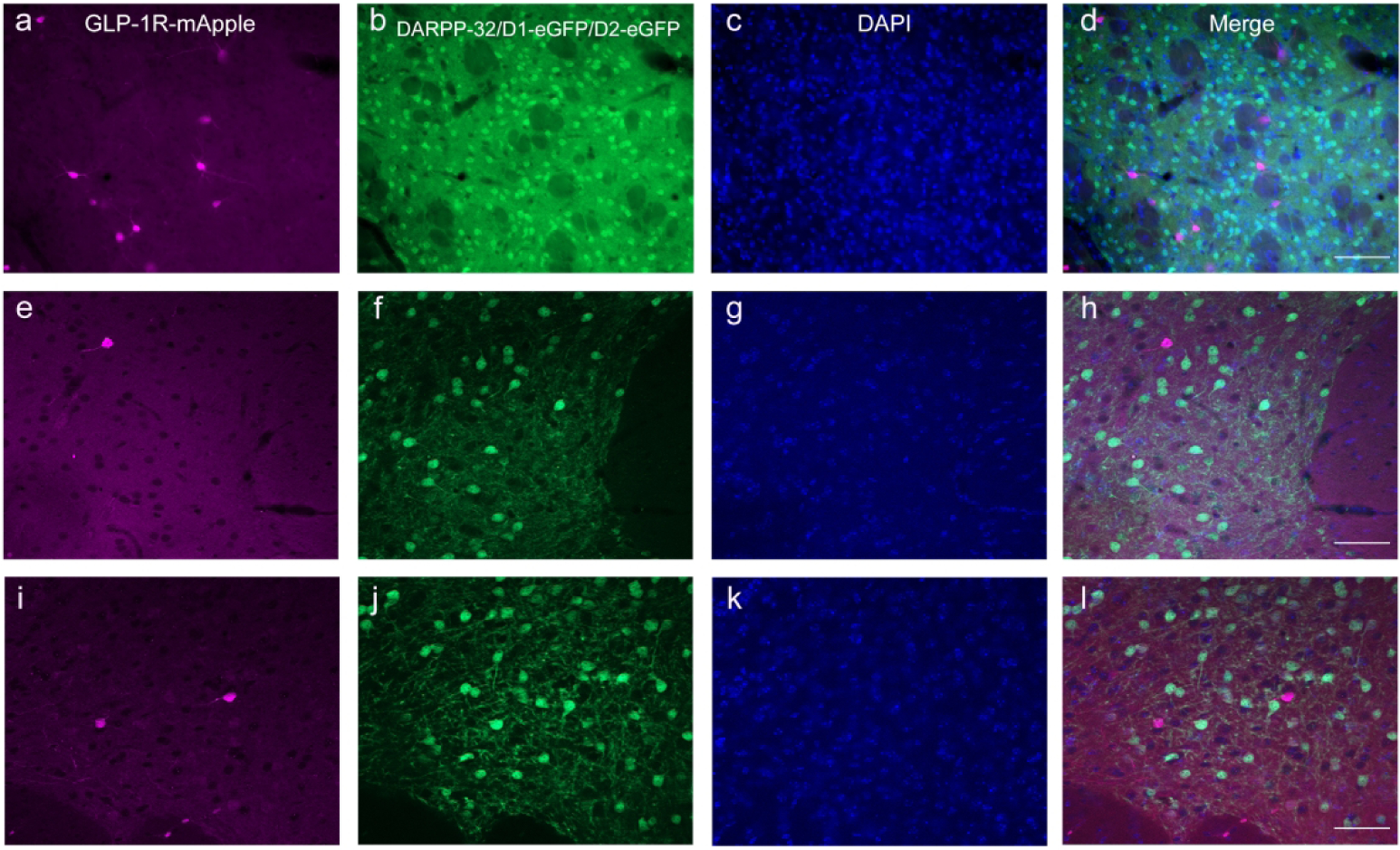
GLP-1R+ cells (a) were not expressed in targets of DAergic projections, as determined by lack of colocalization with DARPP-32 (b, green) within the STR. 10X magnification, 100 μm scale bar. Additionally, there was no colocalization (h, l) of GLP-1R-mApple (e, i) with either the D1R (f) or D2R (j) within the NAc using GLP-1R-mApple mice crossed with either D1- or D2-eGFP mice. 20X magnification, 100 μm (a–d) and 50 μm (e–l) scale bars. DAPI is depicted in blue (c, g, k).