
Figure 8:
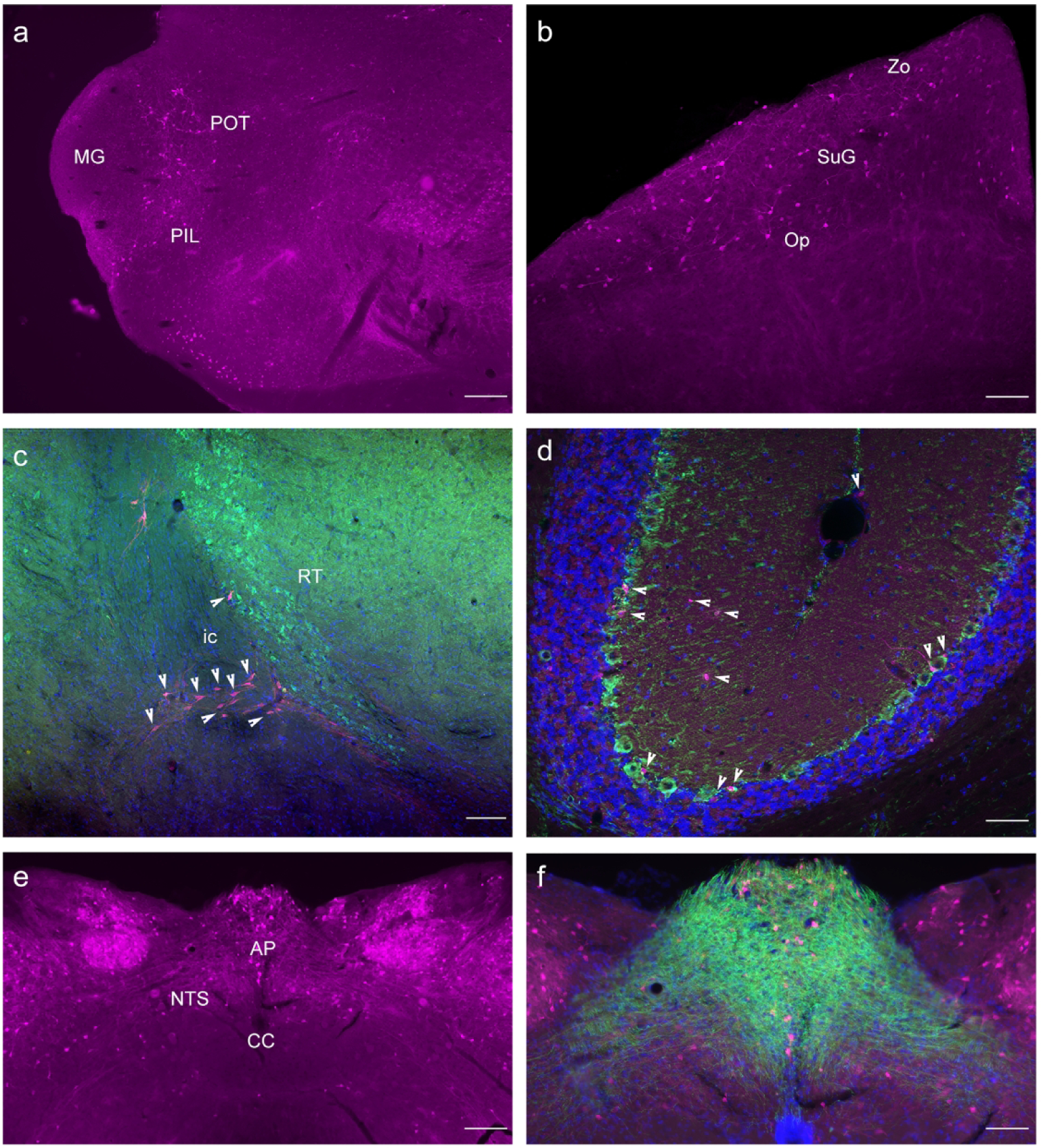
Thalamic expression of GLP-1R-mApple was found within the PoT (a; 5X magnification, 200 μm scale bar) as well as the sc, particularly the superficial gray and optic nerve layers (b, 10X magnification, 100 μm scale bar). GLP-1R-mApple was not present within the Rt, visualized via PV staining (green, c; 10X magnification, 100 μm scale bar). The cerebellum demonstrated little expression of the reporter (d, 20X, 50 μm scale bar; GAD67 in green, DAPI in blue). The NTS showed evidence of GLP-1R expression (e, 5X, 200 μm scale bar) which are surrounded by 5-HT3 receptor+ fiber tracts (f, 10X magnification, 100 μm scale bar).