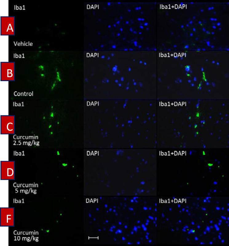
Figure 3.
The effect of curcumin on the activation of spinal microglial cells in morphine-dependent rats. Staining was carried out by using Iba1 (green colored) in the lumbar part of the spinal cord. DAPI staining (blue colored) was used to identify neurons’ nuclei. Magnification ×400; Scale bar, 20 micrometer; Iba1, ionized calcium-binding adapter molecule 1; DAPI (4′,6-diamidino-2-phenylindole); Control, normal saline + DMSO

