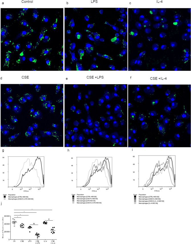
Figure 6.
Effects of CSE, CSE + LPS and CSE + IL-4 on microsphere uptake by MDMs. After 24 h of exposure to CSE, the culture medium was renewed with medium containing fluorescent microspheres (size: 100 nm) and incubated overnight. After incubation, the culture medium was discarded, and the cell monolayers was fixed with paraformaldehyde prior to observation under the confocal microscope (a–f). The fluorescence emitted by microspheres (on channel FL1-H) inside cells was analyzed using CellQuest cytometry software (g–i). The intrinsic FL1-H fluorescence of MDMs was measured in the absence of microspheres. The data correspond to the mean ± SEM of representative experiment of one the 4 donors. *p < 0.05, compared with the control; αp < 0.05, compared with LPS; #p < 0.05, compared with CSE + IL-4.

