Figure 6.
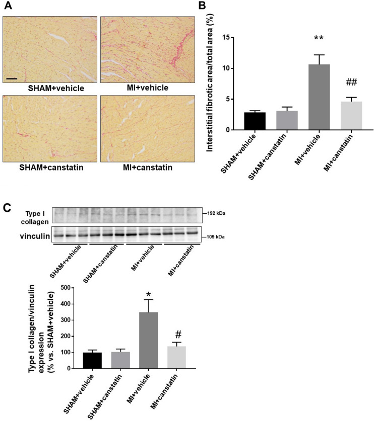
Canstatin prevents interstitial fibrosis in non-infarcted area after MI. Recombinant mouse canstatin (20 μg/kg) or vehicle was intraperitoneally administered for 28 days after LAD ligation. (A, B) The hearts were isolated and then thin paraffin Sects. (4 μm) were made. (A) Representative picrosirius red stained pictures of the non-infarcted areas from SHAM + vehicle, SHAM + canstatin, MI + vehicle and MI + canstatin groups were shown. Scale bar: 100 μm. (B) Interstitial fibrotic area/total area was measured and shown as mean ± S.E.M. (SHAM + vehicle and SHAM + canstatin: n = 6; MI + vehicle and MI + canstatin: n = 8). (C) The tissue protein of non-infarcted area was extracted. Expression of type I collagen was detected by Western blotting. (Upper) Representative blots of type I collagen and vinculin were shown. (Lower) Levels of type I collagen were corrected by vinculin and the normalized expression relative to SHAM + vehicle was shown as mean ± S.E.M. (SHAM + vehicle and SHAM + canstatin: n = 6; MI + vehicle and MI + canstatin: n = 8). *, **P < 0.05, 0.01 vs. SHAM + vehicle, #, ## P < 0.05, 0.01 vs. MI + vehicle (two-way ANOVA followed by Tukey’s post hoc test).

