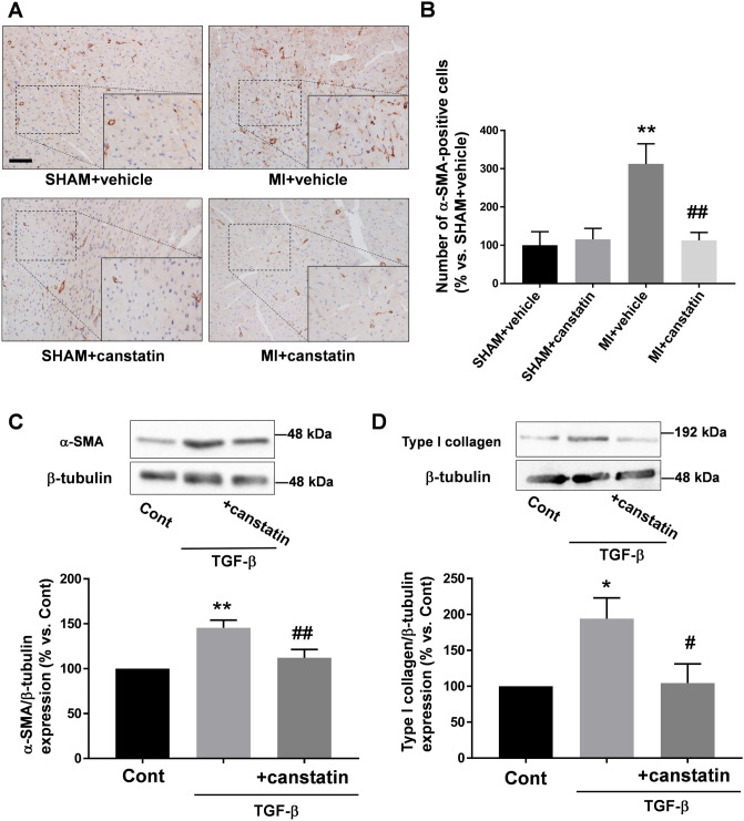
Figure 7.
Canstatin prevents increase of myofibroblasts in non-infarcted area after MI and suppresses transforming growth factor (TGF)-β1 -induced differentiation of cardiac fibroblasts into myofibroblasts. (A, B) Recombinant mouse canstatin (20 μg/kg) or vehicle was intraperitoneally administered for 28 days after LAD ligation. The hearts were isolated and then thin paraffin Sects. (4 μm) were made. (A) Representative pictures of the non-infarcted areas from SHAM + vehicle, SHAM + canstatin, MI + vehicle and MI + canstatin groups reacted with a specific antibody against α-smooth muscle actin (α-SMA) were shown. Scale bar: 100 μm. (B) The number of myofibroblasts, a non-vascular α-SMA-positive cell, in 3 fields was counted, and the normalized number relative to SHAM + vehicle was shown as mean ± S.E.M. (SHAM + vehicle and SHAM + canstatin: n = 6; MI + vehicle and MI + canstatin: n = 8). **P < 0.01 vs. SHAM + vehicle, ## P < 0.01 vs. MI + vehicle (two-way ANOVA followed by Tukey’s post hoc test). (C) Cardiac fibroblasts isolated from ventricles of normal rats were stimulated with TGF-β1 (10 ng/ml) for 48 h in the presence or absence of recombinant mouse canstatin (250 ng/ml, 30 min pre-treatment), and the cell lysates were collected. Expressions of α-SMA (C) and type I collagen (D) were detected by Western blotting. (Upper) Representative blots of α-SMA, type I collagen and β-tubulin were shown. (Lower) Levels of α-SMA and type I collagen were corrected by β-tubulin, and the normalized expressions relative to Cont were shown as mean ± S.E.M. (C: n = 7; D: n = 5). *, **P < 0.05, 0.01 vs. Cont, #, ## P < 0.05, 0.01 vs. TGF-β (one-way ANOVA followed by Tukey’s post hoc test).

